How Do You Know What Is Actually in the Vial?
If a treatment is described as MUSE cell therapy, a general stem cell label is not enough. The question how are MUSE cells isolated is about how a laboratory selects and identifies the population it intends to administer.
This guide follows the selection pathway. It does not repeat the general MSC release-certificate checklist, or laboratory-directed pre-differentiation.
Which Tissue Was Used in the Published Method?
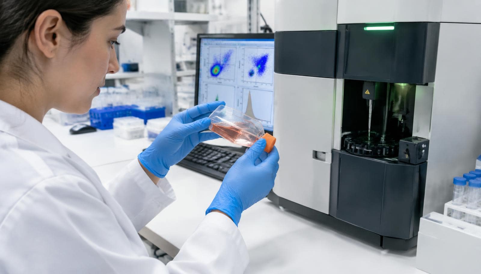
The foundational work identified cells within bone-marrow stromal cultures and skin fibroblasts. Heneidi and colleagues investigated adipose tissue. Dushime and colleagues isolated SSEA-3-positive cells from the Wharton's jelly matrix of umbilical cord tissue by flow cytometry and investigated them in a mouse model of radiation-induced intestinal injury. Each paper's source and processing method matters.
Bone marrow, fat and cord tissue are not interchangeable descriptions. A finding in one source does not establish the purity, growth behaviour or clinical evidence of a preparation from another.
What SSEA-3 Sorting Does
SSEA-3 is a cell-surface marker used in published MUSE identification and selection. Antibody-based sorting can enrich SSEA-3-positive cells from a mixed starting population. Foundational methods also investigated stress-based enrichment.
Enrichment is a selection step, not proof of treatment effectiveness. The report should distinguish marker-positive cells in the starting culture from the proportion in the final preparation, and identify when the measurement was made.
What Expansion Must Be Distinguished From Selection
After isolation, published experiments use defined culture conditions to study expansion and cell behaviour. Multiplying cells does not automatically establish that the final population retains the same identity as the sorted sample.
The laboratory should explain what it measures after culture, rather than relying only on a pre-expansion result. This is a product-specific question, not evidence that every MUSE preparation follows one published manufacturing method.
Comparing Identity With Readiness for Administration
| Question | Information to request | What it cannot prove alone |
|---|---|---|
| Was the intended population selected? | Source, selection method and SSEA-3 analysis | Clinical effectiveness |
| What remains after expansion? | Final identity results and culture history | Safe administration in every patient |
| Was the batch released under its specification? | Viability and contamination results with methods and acceptance limits | Benefit for a particular condition |
A release document should connect the actual batch with source traceability, identity, viable cell count, sterility, mycoplasma and endotoxin information, test dates and release authorisation. Ask which tests are complete and how any pending results are handled. These are manufacturing questions, not a universal validated MUSE protocol.
Montemurro and colleagues' 2024 report describes manufacturing and quality testing of cord-blood MSCs, not isolated MUSE cells or cord-tissue MUSE cells. It provides a general MSC manufacturing example, not a validated MUSE release specification or evidence of MUSE clinical safety.
Our in-house laboratory content describes MRC's cell-processing context. A GMP-standard facility description alone does not demonstrate MUSE-specific identity or equivalence to a clinical-trial product.
MUSE Cell Therapy Approach
- How it works: naturally occurring selected cells are studied for their interactions with injured tissue; isolation is not genetic reprogramming.
- Who may be suitable: patient eligibility requires applicable condition-specific evidence and oversight. A marker result cannot determine candidacy.
- What a session involves: clinical CL2020 reports used intravenous administration with monitoring. A different source or isolation method cannot be assumed to follow that evidence.
- Limits: manufacturing identity does not establish benefit. MSCs, pre-differentiated cells and other regenerative options require separate assessment.
Connect the preparation to the evidence: MUSE stroke study, MUSE heart attack study and tumour and immune safety questions. Neither the tinnitus discussion nor a general MUSE label establishes evidence for another diagnosis.
Four Questions for the Laboratory
- What tissue and selection method produced this batch?
- Which final-product assay demonstrates the intended identity?
- Which dated viability and contamination results authorised release?
- Which published clinical preparation, if any, is genuinely comparable?
If those answers are unclear, resolve them before proceeding. The existing MUSE overview supplies the cell-type background without replacing product documentation.
Related reading: how MUSE cells differ from iPS cells.
Medical References
- Kuroda Y and colleagues, 2010. Unique multipotent cells in adult human mesenchymal cell populations
- Wakao S and colleagues, 2011. Multilineage-differentiating stress-enduring (Muse) cells are a primary source of induced pluripotent stem cells in human fibroblasts
- Heneidi S and colleagues, 2013. Awakened by Cellular Stress: Isolation and Characterization of a Novel Population of Pluripotent Stem Cells Derived from Human Adipose Tissue
- Dushime H and colleagues, 2023. Fetal Muse-based therapy prevents lethal radio-induced gastrointestinal syndrome by intestinal regeneration
- Montemurro T and colleagues, 2024. Off-the-Shelf Cord-Blood Mesenchymal Stromal Cells: Production, Quality Control, and Clinical Use
Talk to a Doctor Before You Decide
Ask for the proposed preparation's identity and source information. We can explain the difference between a general MSC report and MUSE-specific characterisation.
Schedule a Consultation